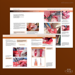
This book, structured according to the normal anatomical areas in radiological diagnostic imaging: abdomen, neck, thorax, limbs, spine, and head, offers the key aspects of both radiographic interpretation and the diagnosis of diseases, as well as a special chapter with the most common diagnostic errors. Its more than 500 high-resolution images are accompanied by the necessary text to increase their descriptive value. In addition, the book is complemented by multimedia material, which can be accessed through QR codes located throughout the text. In this way, the reader can access different normal radiographic anatomy diagrams, both with and without the anatomical details identified. All these elements make this book a reference in the field of clinical radiology.
This book, structured according to the normal anatomical areas in radiological diagnostic imaging: abdomen, neck, thorax, limbs, spine, and head, offers the key aspects of both radiographic interpretation and the diagnosis of diseases, as well as a special chapter with the most common diagnostic errors. Its more than 500 high-resolution images are accompanied by the necessary text to increase their descriptive value. In addition, the book is complemented by multimedia material, which can be accessed through QR codes located throughout the text. In this way, the reader can access different normal radiographic anatomy diagrams, both with and without the anatomical details identified. All these elements make this book a reference in the field of clinical radiology.
Author
Isabel Garcìa Real
She is a Doctor in Veterinary Medicine. She graduated from the Complutense University of Madrid in 1993 and obtained her Ph.D. in 2000. She joined the Department of Animal Medicine and Surgery of the Faculty of Veterinary Medicine of the Complutense University of Madrid in 1993, where she worked as a professor in Radiology.
She has been the Head of the Diagnostic Imaging Serice at the Complutense Clinical Veterinary Hospital since 2005 and Director of the Magnetic Resonance Unit of the hospital since its opening in December 2008. She focuses her educational and research work on radiology, abdominal ultrasound, CT scans, and MRI.
She has worked at the Universities of California (Davis, USA) and Cambridge (UK) and the Animal Medical Center in New York and the Animal Health Trust (Newmarket, UK).
She is the author of several national and international publications and has participated as a speaker in various courses and national and international conferences.
Table of Contents
1. Abdomen
Principles of interpretation
Abdominal wall
Peritoneal cavity and retroperitoneal space
Liver, spleen and lymph nodes
Urinary system
Genital system
Stomach
Small intestine
Large intestine
2. Neck and Thorax
Principles of interpretation
Thoracic wall
Pharynx, larynx and trachea
Oesophagus
Pleural space
Mediastinum
Heart
Lung
3. Appendicular Skeleton
Principles of interpretation
Congenital, hereditary and developmental disorders
Fractures
Tumours and osteomyelitis
Deformations due to disorders of the growth plate
Nutritional and metabolic disorders
Articular pathology
Other disorders of the appendicular skeleton
4. Spine
Principles of interpretation
Myelography
Congenital disorders
Instability syndromes
Degenerative intervertebral disc disease
Other disorders of the spine
5. Head
Principles of interpretation
Cranial vault
Nasal cavity and sinuses
Mandible, maxilar and temporomandibular joint
Teeth
Auditory system
6. Most Common Diagnostic Errors
Abdomen
Neck and thorax
Appendicular skeleton
Spine
Head
Digital image
Data sheet
Specific References
This book, structured according to the normal anatomical areas in radiological diagnostic imaging: abdomen, neck, thorax, limbs, spine, and head, offers the key aspects of both radiographic interpretation and the diagnosis of diseases, as well as a special chapter with the most common diagnostic errors. Its more than 500 high-resolution images are accompanied by the necessary text to increase their descriptive value. In addition, the book is complemented by multimedia material, which can be accessed through QR codes located throughout the text. In this way, the reader can access different normal radiographic anatomy diagrams, both with and without the anatomical details identified. All these elements make this book a reference in the field of clinical radiology.