- On sale!


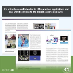
The previous editions of this manual have enjoyed great appreciation; the didactic effectiveness, the clarity and coherence of the book have made it a reference for many students, as well as a practical guide for professionals and specialists. For this third edition, the majority of the chapters have been rewritten and new clinical cases have been added. The book is based on lavishly illustrated clinical cases. Also, given their frequency in oral surgery, proper space at the end of the book is given to the most common post-surgical complications, strategies of prevention, and management.
The first edition of this book was published in 2001 and, now in its third edition, the book has retained all the characteristics that make it a pillar of Oral Surgery. It is extremely clear in describing the basic concepts of oral surgery. Surgical anatomy is described with the support of illustrations whose quality is unmatched in other books. It offers a detailed step-by-step explanation of all the surgical procedures presented. All the new diagnostic and therapeutic procedures, the new materials, and the new surgical techniques find a place in the book. Large space is given to implantology given the importance this discipline has acquired in clinical settings. The book is based on lavishly illustrated clinical cases. Also, given their frequency in oral surgery, proper space at the end of the book is given to the most common post-surgical complications, strategies of prevention, and management.
Strengths:
- Clarity of explanation for beginners to easily grasp the basics of the discipline
- Step-by step description of the Minimally-invasive surgical techniques
- Surgical anatomy is described with the use of extremely high-quality illustrations
- All the new implantology techniques and materials are described
- Correct management and prevention of complications
- Offers several QR codes to digital content related to the topic discussed in each chapter
Author:
Prof. Matteo Chiapasco
Head of the Clinical Unit of Oral Surgery (Department of Biomedical, Surgical and Dental Sciences) of the University of Milan Associate Professor of the University of Milan, Associante Professor of the Loma Linda University in Los Angeles (California – USA)
Table of contents:
Chapter 1 BASIC PRINCIPLES. Introduction. Correct pre-operative evaluation’s steps.
Chapter 2 SURGICAL ANATOMY. Posterior Jaw. Anterior Jaw. Oral Floor. Tongue. Posterior Maxilla. Anterior Maxilla. Palate. Cheek. Upper Lip. Lower Lip.
Chapter 3 FUNDAMEN- TALS OF SURGICAL PROCEDURE Preparation. Local Anesthesia. Incision and flap preparation techniques. Ostectomy. Hemostasis. Sutures.
Chapter 4. DENTAL AVULSIONS. Simple avulsions. Complicated avulsions -open surgery technique.
Chapter 5 DENTAL ELEMENTS. Treatment of dental eruptions. Avulsions. Dental Implants.
Chapter 6 DENTAL INFECTIONS. Pathogenesis. Predisposing Factors. Diagnosis of dental infections. Treatment.
Chapter 7 SURGICAL ENDODONTICS. Visit. Evaluation. Indications. Contraindica- tions. Surgical Treatment. Follow up.
Chapter 8 CYSTS OF THE MAXILLA. Bone Cysts. Cyst of the Maxillary Sinus.
Chapter 9 BENIGN TUMORS OF THE ORAL FLOOR. Introduction. Principle of biopsy. Diagnosis. Treatment.
Chapter 10 SURGICAL PATHOLOGY OF THE SALIVARY GLANDS. Sialolithiasis of Salivary Glands. Cysts and Pseudocysts of Minor Salivary Glands.
Chapter 11 Surgery of the Frenulum and Minor Pre prosthetic Surgery.
Chapter 12 ALVEODENTAL TRAUMATOLOGY. Trauma Classification. Diagnostic eval- uation. Treatment.
Chapter 13 IMPLANTOLOGY Basic Surgical Implantology. Complex Implants Prosthesis.
Chapter 14 COMMON COMPLICATIONS IN ORAL SURGERY. Intraoperative complications. Postoperative complications.
Data sheet
Specific References
The previous editions of this manual have enjoyed great appreciation; the didactic effectiveness, the clarity and coherence of the book have made it a reference for many students, as well as a practical guide for professionals and specialists. For this third edition, the majority of the chapters have been rewritten and new clinical cases have been added. The book is based on lavishly illustrated clinical cases. Also, given their frequency in oral surgery, proper space at the end of the book is given to the most common post-surgical complications, strategies of prevention, and management.