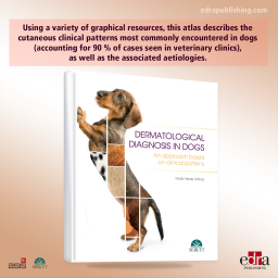
This book describes the most common bacterial skin infections affecting cats and dogs, their clinical presentation, and aetiological agents, besides offering practical techniques and advice for their identification and management using clear images, tables, and diagrams. It also facilitates veterinary surgeons with the tools to treat resistant infections and explains the latest developments in topical and systemic treatments.
This book describes the most common bacterial skin infections affecting cats and dogs, their clinical presentation, and aetiological agents, besides offering practical techniques and advice for their identification and management using clear images, tables, and diagrams. It also facilitates veterinary surgeons with the tools to treat resistant infections and explains the latest developments in topical and systemic treatments.
Author:
Alberto Martin Cordero
Dr Alberto Martin Cordero graduated in animal husbandry and veterinary medicine from the University of Guadalajara (Mexico) and obtained a postgraduate degree in veterinary dermatology at the European School for Advanced Veterinary Studies, University of Luxembourg. He has completed training courses at the NAVC Institute in Florida and internships at the animal dermatology clinics associated with the Colorado State University (California, USA) and Ludwig Maximilian University (Munich, Germany).
He currently devotes his time to private veterinary services and is the owner of Vetderm: Dermatología Veterinaria Especializada, the leading dermatology referral clinic in Guadalajara (Jalisco, Mexico). Dr Martin is an associate professor at the University of Guadalajara’s Department of Veterinary Medicine.
He is a member of the Program Committee for the North American Veterinary Dermatology Forum (NAVDF), a founding member of the Latin American Society of Veterinary Dermatology (SLDV), and a member of the American Academy of Veterinary Dermatology. Furthermore, he is the Latin American representative for the World Association for Veterinary Dermatology (WAVD), area coordinator for veterinary dermatology at the León Veterinary Congress (Mexico), and president of the 4th Latin American Congress of Veterinary Dermatology, 2018 (Mérida, Mexico).
His main interests are diseases of the ear in cats and dogs, allergic skin diseases, and video otoscopy.
He has published several articles and clinical cases in national and international journals, and spoken at conferences in Mexico, Central America, South America, and Eastern Europe.
KEY FEATURES:
➜ Includes practical techniques for the identification and management of infections.
➜ With a great number of images, tables and diagrams
Table of contents:
1. The skin microbiome and diagnosis of bacterial skin infections
2. Superficial pyoderma
3. Deep pyoderma
4. Miscellaneous bacterial infections
5. Bacteriological culture and their interpretation
6. Topical treatments for bacterial skin infections
Data sheet
Specific References
This book describes the most common bacterial skin infections affecting cats and dogs, their clinical presentation, and aetiological agents, besides offering practical techniques and advice for their identification and management using clear images, tables, and diagrams. It also facilitates veterinary surgeons with the tools to treat resistant infections and explains the latest developments in topical and systemic treatments.