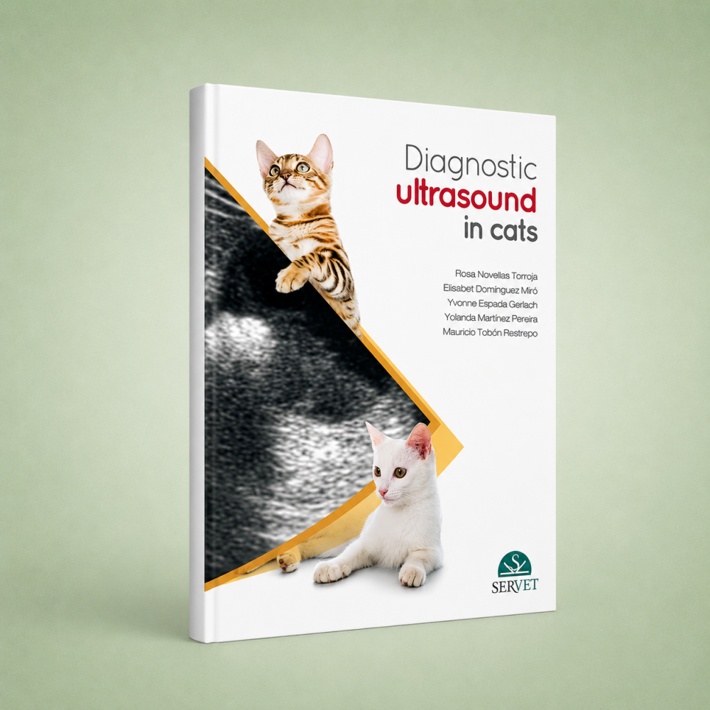


This book provides new insight into feline ultrasound in daily practice. From cranial to caudal, the feline species has been thoroughly scanned, detailing for each body region the scanning technique, as well as the ultrasonography of both the normal and the diseased organ. In each chapter, key points highlight feline species' special features and help the reader focus on the most important. Several short ultrasound clips are also available in the electronic version of the book, increasing, even more, the interest of this book: an up-to-date text and several references to the latest studies.
This book provides new insight into feline ultrasound in daily practice. From cranial to caudal, the feline species has been thoroughly scanned, detailing for each body region the scanning technique, as well as the ultrasonography of both the normal and the diseased organ. In each chapter, key points highlight feline species' special features and help the reader focus on the most important. Several short ultrasound clips are also available in the electronic version of the book, increasing, even more, the interest of this book: an up-to-date text and several references to the latest studies.
Authors:
Rosa Novellas Torroja
I graduated in 2003 from the Universitat Autónoma de Barcelona. After graduating, she developed her Ph.D. project in diagnostic imaging and, in 2007, obtained the Ph.D. at the same university. In 2007, she enrolled a residency training program in veterinary diagnostic imaging at the Royal (Dick) School of Veterinary Studies in Edinburgh. In 2008 she moved to Glasgow, where she continued and finished the residency at Glasgow University Vet School. In 2010 she became a diplomate of the European College of Veterinary Diagnostic Imaging (ECVDI). She is currently working as an associate lecturer at the Universitat Autónoma de Barcelona and a member of the Fundació Hospital Clinic Veterinary diagnostic imaging service.
Elisabet Domínguez Miño
Elisabet graduated in 2005 from the Universitat Autònoma de Barcelona. She subsequently completed an internship in small animal medicine and surgery at the same university. In 2007 she started a Ph.D. project in laboratory animal diagnostic imaging and obtained the Ph.D. in 2011. In 2011 she enrolled a residency training program in veterinary diagnostic imaging at the Fundació Hospital Clínic Veterinary in Barcelona, which she is currently finishing.
Yolanda Martínez Pereira
Yolanda obtained her degree in Veterinary Medicine in 1998 in Spain, and after a period in general practice, she completed an internship in cardiology, obtaining the RCVS Certificate in Veterinary Cardiology (2004). From 2005 to 2008, she completed a residency in Cardiopulmonary Medicine at the University of Edinburgh, followed by a year in the Cardiology Service at the University of Liverpool as a clinician and teacher. During 2009-2012, she joined Borders Veterinary Cardiology Ltd, providing a mobile cardiology referral service in Scotland. She returned to the Royal (Dick) School for Veterinary Studies in 2013 as a lecturer in Cardiopulmonary Medicine, heading the service. She obtained her European Diploma in Veterinary Cardiology in 2009 and became RCVS Specialist in Veterinary Cardiology in 2011.
Yvonne Espada Gerlach
In 1984 she graduated from the Universidad de Zaragoza. After graduating, she developed her Ph.D. project and obtained the Ph.D. in 1990 from the Universitat Autònoma de Barcelona (UAB). She began to work in diagnostic imaging in 1990, performing training in several Diagnostic Imaging Services since then, in Paris (Alfort), United Kingdom (Royal Veterinary College and Animal Health Trust), Germany (Giessen), and in the United States (Virginia Tech University). She became a lecturer of the Animal Medicine and Surgery Department of the Universitat Autónoma de Barcelona in 1991, where she is still working as a lecturer currently. In 1992 she started working in the Diagnostic Imaging Service of the Fundació Hospital Clinic Veterinary (UAB), the Head of the service since then.
Mauricio Tobón Restrepo
In 2006 Mauricio Tobón Restrepo obtained his Veterinary Degree from the Universidad de Antioquia (Medellín, Colombia). Immediately afterward, he worked as a professor of Veterinary Anatomy at the same university. At the same time, he was working in private practice. In 2008, he was employed by the Corporación Universitaria Lasallista as a full-time professor and director of the veterinary laboratory of anatomy. In 2010, he received his Masters in Education. In 2011 he was given a grant from the Colombian government and the Corporación Universitaria Lasallista to study abroad for his Ph.D. studies. He is finishing his doctoral studies in Animal Medicine and Health at the Universidad Autónoma de Barcelona (Spain) to enroll afterward as an ECVDI (European College of Veterinary Diagnostic Imaging) resident at the University of Utrecht (The Netherlands). His research line is on feline diagnostic imaging.
Table of Contents:
1. Ultrasound of the head and neck
Scanning technique
Ultrasound of the normal head and neck
Ultrasound in the study of diseases of the head and neck
2. Echocardiography
Scanning technique
Ultrasound of the normal heart
Ultrasound in disorders of the heart
3. Non-cardiac thoracic ultrasound
Scanning technique
Normal non-cardiac thoracic ultrasound
Ultrasound in thoracic lesions
4. Ultrasound of the liver and biliary system
Scanning technique
Ultrasound of the normal liver and biliary system
Ultrasound in liver and biliary diseases
5. Ultrasound of the gastrointestinal tract
Scanning technique
Ultrasound of the normal gastrointestinal tract
Ultrasound in gastrointestinal tract disorders
6. Ultrasound of the pancreas
Scanning technique
Ultrasound of the normal pancreas
Ultrasound in pancreatic disorders
7. Ultrasound of the spleen
Scanning technique
Ultrasound of the normal spleen
Ultrasound in splenic disorders
8. Ultrasound of the adrenal glands
Scanning technique
Ultrasound of the normal adrenal glands
Ultrasound in adrenal disorders
9. Ultrasound of the kidneys and ureters
Scanning technique
Ultrasound of the normal kidneys and ureters
Ultrasound in renal and ureteral disorders
10. Ultrasound of the urinary bladder and urethra
Scanning technique
Ultrasound of the normal bladder and urethra
Ultrasound in bladder and urethral disorders
11. Ultrasound of the reproductive system
Scanning technique
Ultrasound of the normal reproductive system
Ultrasound in disorders of the reproductive system
12. Ultrasound of the abdominal cavity, lymph nodes and large vessels
Scanning technique
Ultrasound of the normal abdominal cavity, lymph nodes and large vessels
Ultrasound in abdominal cavity, lymph node and large vessel lesions
13. Ultrasound of the musculoskeletal system and superficial soft tissues
Scanning technique
Ultrasound of the normal musculoskeletal
system and superficial soft tissues
Ultrasound in alterations of the musculoskeletal system and superficial soft tissues
14. Sampling and other ultrasoundguided procedures
General considerations and indications
Materials
Technique
Considerations for sampling and other ultrasound-guided procedures
Data sheet
Specific References
This book provides new insight into feline ultrasound in daily practice. From cranial to caudal, the feline species has been thoroughly scanned, detailing for each body region the scanning technique, as well as the ultrasonography of both the normal and the diseased organ. In each chapter, key points highlight feline species' special features and help the reader focus on the most important. Several short ultrasound clips are also available in the electronic version of the book, increasing, even more, the interest of this book: an up-to-date text and several references to the latest studies.