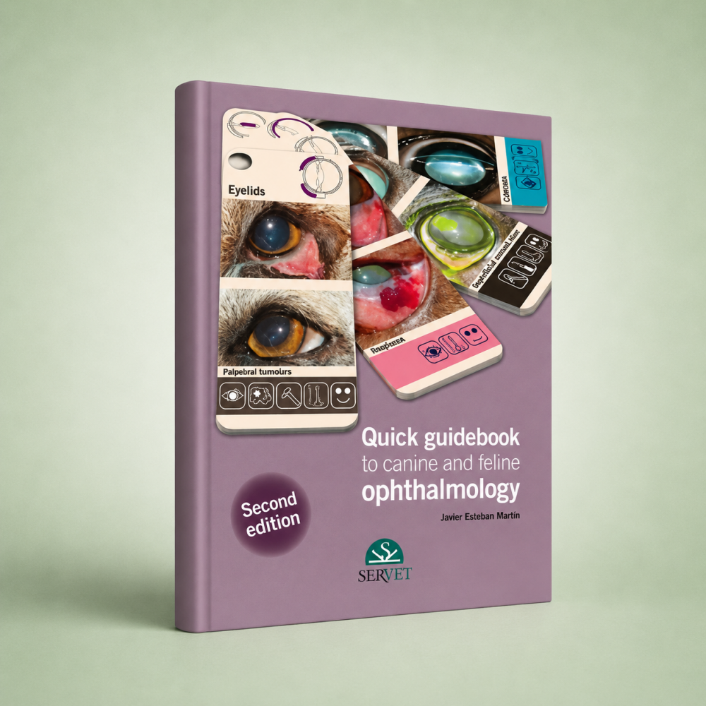


The aim of this guide is to provide both the vet and the student with a quick reference guide to enable them to easily recognize the most common ophthalmological ailments seen in daily practice. More than 700 images from real cases to easily recognize the most common ophthalmological ailments seen in daily practice.
The aim of this guide is to provide both the vet and the student with a quick reference guide to enable them to easily recognize the most common ophthalmological ailments seen in daily practice. More than 700 images from real cases to easily recognize the most common ophthalmological ailments seen in daily practice.
Javier Esteban Martin
Professor of veterinary medicine and clinical surgery, Professor of ophthalmology, School of Veterinary Medicine, University of Murcia, Spain.
Diploma in ophthalmology from the Autonomous University of Barcelona. French certificate of veterinary ophthalmology.Member of the Latin American College of Veterinary Ophthalmologists (CLOVE).
Table of Contents:
Organisation of this guidebook
How to use this guidebook
Key for the Quick guidebook to canine and feline ophthalmology
1 Anatomical structures
Diagram of the eye and its adnexa
Lacrimal apparatus diagram
2 Eyelids
Palpebral agenesis
Palpebral dermoid
Entropion in a Shar Pei
Entropion in a Chow Chow
Entropion (surgical techniques)
Medial entropion
Lateral entropion
Cicatricial entropion
Entropion due to lack of support
Ectropion
Entropion/ectropion
Entropion/ectropion. Diamond eye
Euryblepharon
Palpebral laceration
Fungal blepharitis
Bacterial blepharitis
Parasitic blepharitis
Blepharitis (hypersensitivity)
Immune blepharitis
Palpebral neoplasms
Distichiasis
Districhiasis
Ectopic cilia
Trichiasis
Trichomegaly
Palpebral ptosis
Lagophthalmos
Chalazion
Hordeolum
Lentigo
Foreign bodies
3 Nictitating membrane
Prolapse of the nictitating membrane gland
Cartilage eversion
Plasmoma
Follicular conjunctivitis
Granulomas due to Leishmania
Lacerations/tears
Protrusion of the nictitating membrane
Neoplasms
Foreign bodies
Depigmentation of the free edge of the nictitating membrane
4 Conjunctiva
Conjunctival dermoid
Conjunctival cysts
Conjunctivitis
Keratoconjunctivitis sicca
Symblepharon
Drug plaques
Mucinosis
Haemorrhages
Chemosis
Wounds
Neoplasms
5 Cornea and sclera
Superficial corneal ulcer
Refractory corneal ulcer
Stromal ulcer
Deep corneal ulcer
Descemetocoele
Corneal perforation
Melting corneal ulcers
Chronic superficial keratitis
Keratoconjunctivitis sicca
Pigmentary keratitis
Feline eosinophilic keratitis
Corneal dystrophy
Corneal degeneration
Corneal oedema
Neoplasms
Foreign bodies
Feline corneal sequestrum
Dermoid cyst
Corneal abscess
Episcleritis
6 Uveal tract
Iris hypoplasia
Iris heterochromia
Persistent pupillary membrane
Uveitis
Iris cysts
Neoplasms
Iris atrophy
Melanosis
Hyphaema
7 The lens
Microphakia
Coloboma
Nuclear sclerosis
Cataracts
Lens subluxation
Lens luxation
8 Ocular fundus
Normal ocular fundus
Retinal detachment
Retinal haemorrhage
Retinal dysplasia
Optic nerve hypoplasia
Optic neuritis
Retinal degeneration
9 Globe and orbit
Enophthalmos
Exophthalmos
Atrophy of the globe
Buphthalmos
Retrobulbar abscess
Orbital cellulitis
Orbital neoplasms
Foreign bodies
Proptosis
10 Lacrimal apparatus
Epiphora
Keratoconjunctivitis sicca
Imperforate lacrimal punctum
Hypoplasia
Obstruction
Dacryocystitis
11 Glaucoma
Diagnosis
Classification
Clinical signs
Differential diagnosis
Treatment
Sequelae
Bibliography
Alphabetical index
Data sheet
Specific References
The aim of this guide is to provide both the vet and the student with a quick reference guide to enable them to easily recognize the most common ophthalmological ailments seen in daily practice. More than 700 images from real cases to easily recognize the most common ophthalmological ailments seen in daily practice.