
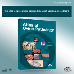
Atlas of Ovine Pathology
Price
$162.79
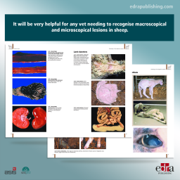
This atlas compiles clinical cases and images of pathological conditions. It will be very helpful for any vet needing to recognise macroscopical and microscopical lesions in sheep.