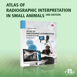


This second edition of the book enhances its original goal of aiding veterinary practitioners in radiographic interpretation and pathological diagnosis. It builds upon the first edition by introducing a chapter on radiology principles and a comprehensive atlas for radiographic positioning and anatomy. The edition includes 400 new images and has updated existing graphics to improve quality. Additionally, the book features new multimedia content accessible via QR codes in the text, allowing readers to view diagrams of normal radiographic anatomy, both labeled and unlabeled, for better understanding and reference.
This second edition extends its initial aim of assisting general veterinary practitioners in radiographic interpretation and pathological diagnosis by adding to the content of the first edition a chapter on the principles of radiology and a complete atlas of radiographic positioning and anatomy. It includes 400 new images and updates previous graphic material content to further increase its quality. In addition, the book is complemented by new multimedia material, which can be accessed through QR codes located throughout the text. In this way, the reader can access different normal radiographic anatomy diagrams, both with and without the anatomical details identified.
Data sheet
Specific References