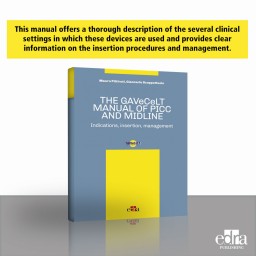
The GAVeCeLT manual of Picc and Midline
The use of intravenous access devices is fundamental for all patients needing frequent blood sample collection, artificial nutrition, chemotherapy, antibiotic therapy, and any other intravenous treatment.
Active filters
The use of intravenous access devices is fundamental for all patients needing frequent blood sample collection, artificial nutrition, chemotherapy, antibiotic therapy, and any other intravenous treatment.
In 23 chapters, the treatment of pediatric patients is examined in every aspect: from the approach to the patient, to prevention, to conservative treatment in carious pathology. A review of the endodontics and pharmacotherapy are some of the available updates, which complete the comprehensive overview of the discipline, along with the new chapters on miofunctional therapy, on the dental treatment of snoring and nocturnal apnoea syndrome, on the treatment of patients with disabilities.
This illustrated work has the aim to assist the veterinary surgeon with his/her communication with the pet owner. Following the last book spirit dedicated to the surgery, in this atlas, the drawings about internal and external parasites of dogs and cats will make easier the explanations of the veterinary surgeon to his/her customers, as well as the control, prevention, and treatment plans that the veterinary surgeon needs to set up in some cases. The clarity and accuracy of the drawings, designed to make understandable and to reduce the time spent on the explanations to the pet owner, make this atlas another useful clinical tool. This book is aimed at veterinary surgeons, students, teachers, and professionals in this field.
Servet presents the third volume of the publication “Small animal surgery”, which describes the main surgical procedures on the cranial abdomen of the dog, cat and ferret. This work, which has been acclaimed by professionals, stands out for the excellence of its images and the step-by-step description of each surgical technique using real clinical cases.
Leukemia is a heterogeneous group of lymphohematopoietic neoplastic diseases affecting cats and dogs with a relatively high frequency and generally showing non-specific clinical signs. A correct diagnosis of the different types of leukemia (Acute Lymphoid Leukemia, Acute Myeloid Leukemia, Chronic Lymphocytic Leukemia) and the identification of putative prognostic factors are based on the use of different laboratory tests including hematology, cytology, bone marrow biopsy, flow cytometry/immunohistochemistry. A comprehensive interpretation of the laboratory results is mandatory to differentiate leukemia from leukemic lymphoma and other similar hematological diseases, accurately identify the type of leukemia, and adequately predict prognosis and tailor therapy.
Nephropathology is a unique specialization in anatomic pathology, and a complete set of analyses comprising LM, IF, and TEM should always be considered. When performing a renal biopsy, few criteria should be encountered. First, the biopsy should be considered a procedure to improve therapy, and the biopsy should be done safely. An expert and specialized neuropathologist should evaluate the tissue sample to get the most informative description using all the methods required to characterize the pathological changes. The required methods for renal biopsy evaluation include special staining protocols for light microscopic (LM) evaluation, as well as for transmission electron microscopic (TEM) and immunofluorescence evaluations (IF).
This practical text on small animal dermatology provides the knowledge necessary to successfully diagnose and treat the most common skin diseases, which can account for up to 90% of veterinary consultations. This information is presented together with numerous images as well as a large number of diagnostic algorithms, which add to the practical value of the book.
Diagnostic imaging is a key component in the care of companion animal patients. In recent decades the available imaging modalities have expanded from conventional radiography to include ultrasonography, computed tomography, and magnetic resonance imaging, and all are employed on a daily basis in both general and specialty veterinary practice.
This is not the ordinary academic text to have in your library. It’s the result of the commitment of the Author, a practicing clinician who has focused his career on practical solutions to treat thousands of cases.
The ultimate objective of root canal therapy is the three-dimensional obturation of the endodontic space after it has been completely cleaned, shaped, and disinfected.
The purpose of obturation is to seal all "portals of exit" to impede any sort of communication or exchange between the endodontium and periodontium.
First release 1.1.2023 - Available until 12.31.2023
Two canals in the same root could each have their apical foramen or converge into a single foramen. This characteristic could apply to any root containing two canals internally. Still, it is crucial to remember this when preparing the mesial canals of lower molars and mesiobuccal canals of upper molars.
The presence of two canals with separate foramen does not require any particular know-how during the preparation phase but only in the obturation phase.
When the two canals converge in a common foramen instead, it is necessary to take some precautions during the shaping phase and ascertain the presence of this anatomic peculiarity as soon as possible.
First release 1.1.2023 - Available until 12.31.2023
Perforations are pathological or iatrogenic communications between the root canal system and the attachment apparatus. The clinician must take special care to avoid tooth perforations during endodontic therapy, as perforation requires additional treatment.
First release 1.1.2023 - Available until 12.31.2023
Occasionally, a nonsurgical root canal procedure won't be enough to save a tooth, and endodontic surgery will be recommended. Advanced technologies like digital imaging and operating microscopes allow these procedures to be performed quickly, comfortably, and successfully.
First release 1.1.2023 - Available until 12.31.2023
Sometimes, a nonsurgical root canal procedure is not enough to save a tooth, and endodontic surgery is recommended.
Techniques, materials, and knowledge are essential to make microsurgical endodontics approaches accurate and predictable in treating endodontic lesions.
First release 1.1.2023 - Available until 12.31.2023
For years, two-dimensional (2D) radiographic examinations, dental panoramic, and periapical radiographs have been used.
The interpretation of a two-dimensional image can be complicated by several factors, including the anatomical area and the overlap of other teeth and surrounding dentoalveolar structures.
These problems can be overcome by using images obtained with small or otherwise limited volumes of Cone Beam Computed Tomography, which provides accurate three-dimensional images of the teeth and surrounding dentoalveolar structures. CBCT gives dentists the advantage of being able to carry out correct diagnoses and, above all, correct treatment plans.
First release 1.1.2023 - Available until 12.31.2023
The use of MTA in nonsurgical endodontics: direct pulp capping and treatment of immature apices with the “Apical Barrier Technique”
In the late 90s, Dr. Mahmoud Torabinejad of Loma Linda University, California, developed a new cement named Mineral Trioxide Aggregate which appears to have all of the characteristics requested of the ideal cement to seal pathways of communications between the pulp and the oral cavity (mechanical and carious pulp exposures), and between the root canal system and the periodontium (iatrogenic perforations, open apices, resorbed apices, root-end preparations).
First release 1.1.2023 - Available until 12.31.2023
Successful endodontic treatment depends on completely removing the contents of the root canal system and sealing all communication pathways between the endodontium and periodontium. In other words, the result of endodontic treatment must be the same as obtained with the extraction of a compromised tooth.
First release 1.1.2023 - Available until 12.31.2023
The extraordinary characteristic of super elasticity and strength of the NiTi alloy has made it possible to produce rotary instruments capable of assuring a perfect shaping reproducible with the use of very few instruments, in a short time, without the need for the operator to have the above average manual skills.
First release 1.1.2023 - Available until 12.31.2023
The new ProTaper Next NiTi rotary files represent the latest generation of NiTi rotary files. Thanks to this new tool, we can now more easily achieve the shape of the root canal after preliminary canal enlargement and negotiation of the glide path.
First release 1.1.2023 - Available until 12.31.2023
This class features ten comprehensive webinars to walk clinicians through the complexities of endodontics, from an accurate diagnosis to the best treatments. Several topics are covered, from root canal anatomy, pathologies, radiographs, and scans, to the latest therapeutic endodontics materials, tools, and equipment. Each topic is presented through a review of actual cases and a thorough analysis of scientific literature.
First release 1.1.2023 - Available until 12.31.2023
A new application method and a different clinical reasoning compared to other types of taping and bandaging are the basis of the NeuroMuscular decompressive and compressive taping technique. To illustrate this innovative rehabilitation treatment, this book aims to answer these fundamental questions: When is NeuroMuscular Taping applied? How is it applied? What clinical advantages does it offer?
Although nasal cytology is still a little used diagnostic approach, it has often proved to be decisive: it makes it possible to distinguish between different forms of rhinopathy – inflammatory from infectious, allergic from non-allergic vasomotor, and bacterial from viral and fungal forms, as well as to diagnose the presence, in the same patient, of various “overlapping” forms.
Diseases of the liver and pancreas can cause a degree of despair in veterinary practitioners: so much about the causes and treatments of these diseases is poorly understood, and too many cases remain idiopathic. This book guides both first and second opinion practitioners through clinical assessments and workups, preventing oversight of potential underlying causes. Subsequent sections meticulously delve into pancreatic and liver diseases, encompassing clinical pathology, imaging, biopsy acquisition, cytology, and histology, all expertly elucidated by specialists.